Longitudinal and transverse section photos were photograph with a microscope combined with a digital camera.
19 longitudinal section photos and 43 transverse sections photos from four blocks were taken at 1,5 lens, and 0,5 zoom lens. 17 close-up of organs were taken at 2,5 lens and 2,5 or 1,6 zoom lens. Few longitudinal sections were bigger than the field of the camera, so they were taken in two goes.
Digital acquisition format was jpeg.
Animal selection
Medaka fishes were provided by Dr. M. EDERY, INSERM research director, researcher at USM 505 « Ecosystèmes et interactions toxiques » at the Natural History National Museum (Muséum National d’Histoire naturelle), responsible for the animal facilities of this unit.
In order to obtain whole
animals on longitudinal section slides, two months old animals were chosen.
At this age, they are still small but already sexually mature.
Two males and two females were chosen according to secondary sexual characters
for the atlas . One fish of each sex was used for the longitudinal or transverse
sections.
Aneasthesia and fixation
Fishes were anaesthetized in iced water
during few minutes and then were placed in 4°C cold Bouin’s fluid
for 48 hours. The tail was cut just before fixation in order to permit a
better absorption of the fixator.
Bouin fluid was chosen as a fixator, because it softens bones structures and
avoids the decalcification step.
Embedding and sectioning
Paraffin wax embedding follows three steps:
- sample dehydratation
- impregnation in liquid paraffin wax
- paraffin blocks formation
Samples are first dehydrated in alcohol
and butanol baths. After 48h, they are taken out of the fixator, rinsed in
water and in 50° ethanol. Then they are put for two days in 70°
ethanol at 4°C with bath changed twice to three times a day until samples
have lost the yellow color of the Bouin’s fluid. Then they are put
in 90° ethanol for 24h at 4°C and transfered in butanol in which
they are stocked until embedding.
Fishes are then placed in 56°C molten wax for embedding.
Manuel embedding in 56°C paraffin wax ( wax was changed every day for a
week) has been preferred to vacuum embedding techniques which can damage
fish tissues. Then fishes are put in a mould containing molten wax. For longitudinal
sections fishes are arranged on side on the bottom of the mould. For transverse
sections a cut behind gills is performed. Obtained pieces are arranged vertically
in the mould. Moulds are then allowed to cool and harden. Obtained blocks
are stocked at 4°C.
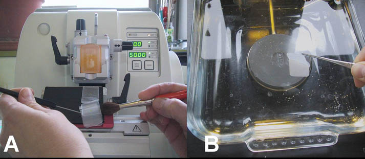
3.5 µm slices were obtained
from the blocks on a Leica ® RM2245 microtome and placed on superfrost
® glass slides using a water bath (see photo) for staining. Every 100
µm, three transverse sections and two longitudinal consecutive sections
were placed on slides.
After drying at 37°C during at least 2 hours, slides were stained in HES (Hematoxylin-Eosin-Safran), standard staining in histology. Hemotoxylin stains nucleus in purple, eosin, cytoplasm in pink and saffron stains in orange collagen fibers.
This staining was performed thanks to a staining-automaton Leica ® CV5030/ST5020 or manually according to the following protocol:
- Three 15 minutes toluene baths
- Two one minute 100° alcohol baths
- One one minute 95° alcohol bath
- Rinsing in four tap water baths
- One 2 minutes hematoxylin bath
- Four distilled water baths
- Short soaking in a hydrochloric acid bath (HCl)
- Water rinsing
- One eosin bath of one minute
- Four water rinsing baths
- Short soaking in one 100° alcohol bath
- One saffron bath (5 minutes)
- Short soaking in two alcohol 100° baths
- Three toluene baths of 2 minutes
- Slides mounting with Eukitt ®